| Expand | ||
|---|---|---|
| ||
Transmission IRM is recommended where possible and can be is predominantly used for solid , samples. If you’re interested in liquid or gaseous samples, please contact the IRM team. Transmission requires that samples be very thin (between 5-10 µm), flat and highly polished where applicable. Solid samples should be microtomed to the correct thickness ranged for analysis, either freestanding or deposited onto an IR transmitting window such as CaF2, BaF2, ZnSe (preferably 0.5 mm in thickness) depending on the window properties and your required wavenumber coverage. Less frequently, Si or silicon nitride (Si3N4) may be used as substrates for transmission IR. A micro-compression cell with diamond or CaF2 windows, to sandwich your sample in place under the beam, is also available for use. Examples of samples for transmission:
|
| Expand | ||
|---|---|---|
| ||
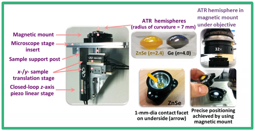
ATR relies on a form of internal reflection to enable the analysis of more difficult samples with IR spectroscopy, e.g. samples that cannot be microtomed for transmission measurements or that do not adequately reflect IR for reflection. Compared to transmission and reflectance FTIR, ATR offers the advantage of enhanced spatial resolution (where a crystal with a high refractive index is used such as Ge). Furthermore, the total internal reflectance phenomenon that ATR relies on (occurs when IR radiation travels through a high refractive index crystal a onto a low refractive index sample surface), results in an evanescent wave that penetrates the surface of the sample, rather than the bulk The IRM beamline has a Bruker single-bounce micro-ATR accessory with a Germanium crystal tip (diameter=100 microns, refractive index=4). Traditional microscopic ATR methods often cause sample damage, contamination and re-positioning issues, where the ATR crystal is attached to the objective. However, with the macro-ATR methods developed in-house and described below, these issues are avoided by single contact ATR mapping. Two models of the macro-ATR are available for use: “hybrid” macro-ATR
Examples of samples for hybrid macro-ATR:
If you are interested in using the macro-ATR acquisition mode, please read the instructions on sample preparation HERE 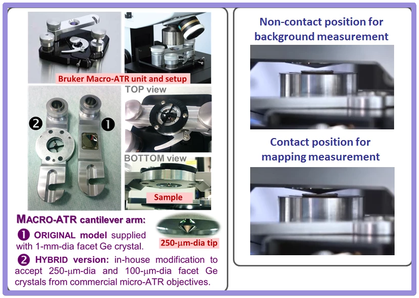
 |
...
| Expand | ||
|---|---|---|
| ||
The IRM beamline has a Bruker Vis/IR Grazing Angle Objective (GAO, 15×) for grazing incidence analyses. This objective is specifically designed to analyse thin layers or films <1 µm thick that are deposited onto non-IR absorbing, reflecting surfaces e.g. highly polished metal slides (Au or Al) or indium tin oxide (ITO) coated glass, for example. GAOs increase the optical path length of a sample by essentially skimming the beam across the surface using high angles of incidence. This objective uses an incident angle of ~84° and shows excellent sensitivity down to monolayer thicknesses, particularly when used with polarised light, and good spatial resolution. The Bruker design relies on transflectance from the sample surface, effectively increasing the sample path length to again assist sensitivity, and uses a polariser in the IR beam; p-polarised light is polarised perpendicular to the surface of the sample and shows enhanced IR absorption when large angles of incidence are used. 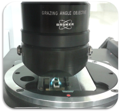  Examples of samples grazing angle experiments:
|
| Expand | ||
|---|---|---|
| ||
Time resolved spectroscopy (TRS) using a FTIR spectrometer offers the advantage of being able to monitor rapidly changing chemical or physical properties of a system in high temporal resolution (up to 65 spectra/sec using a 160 kHz scan velocity @ 16 cm-1). It allows for the simultaneous observation of the progression and/or decay of various chemical species during an event. The rapid scan experiments can be initiated by an external trigger, such as a change in voltage, which can be programmed into the rapid-scan method editor in OPUS, making it ideal for the observation of kinetic chemical changed induced by an electrochemical potential event, to provide an example, More information on rapid scan FTIR spectroscopy can be found on the Bruker website. |